The Modelling & Simulation Community
The Advantages of Membership
NAFEMS is the only worldwide independent association dedicated to engineering modelling, analysis & simulation.
It has been our mission since 1983 to facilitate and promote the safe and efficient use of engineering simulation and analysis.
We're non-profit. We're independent. We're the voice of the CAE community.
Membership has many advantages

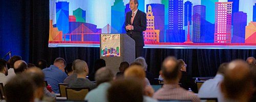
NAFEMS World Congress 2025
The largest independent, international conference dedicated exclusively to engineering simulation.
Attendees discovered new technologies & innovative techniques, whilst networking with end-users, software vendors, consultants and academics, in this worldwide celebration of simulation.

NAFEMS Training
Traditional methods of training are changing. NAFEMS is stepping up to the challenge by providing you with a range of training options, all available remotely, all available from home, and all keeping to the same, internationally renowned, independent standards.

NAFEMS Events
NAFEMS provides the engineering analysis community with as many as fifty seminars, conferences, workshops and open forums throughout the world each year. As the only truly independent organisation dedicated to engineering analysis and simulation NAFEMS' events are widely held to encompass the broadest and most accurate view of the community available.

Your Community
This is your community. Now more than ever, we're working harder to deliver more opportunities for you to stay connected during these challenging times.
Our webinar programme has been stepped up, we're introducing new more interactive online sessions, and our working groups are opening up some of their meetings to the wider community as we come together as one.
You can also continue to submit articles for BENCHMARK Magazine, as well as taking a look at our invitations to tender for new books and documents for the analysis community.
Do you have an idea you want to share? Leave us some feedback.
Resources
The NAFEMS Resource Centre provides a gateway to all of the technical material produced by NAFEMS. It enables access to our publications, magazine articles, webinars, papers and presentations.
Relevant material can be found by searching by keyword, filtering by multilevel technical categories or filtering by author, organisation, or type of resource.
There are currently over 8,000 resources in the centre with this number growing weekly.

Professional Development
As an organisation dedicated to promoting the best use of simulation and analysis and with our wealth of experience within the analysis and simulation industry, there is no better place to further your professional development with our quality and worthwhile training to reach your goals.
Keep track of your development using the PSE Competency Tracker, and get PSE Certified to demonstrate your skills to current and future employers.

What is NAFEMS?
NAFEMS is the International Association for the Engineering Modelling, Analysis and Simulation Community.
We are a not-for-profit organisation which was established in 1983.
Our principal aims are to:
Improve the professional status of all persons engaged in the use of engineering simulation
Establish best practice in engineering simulation
Provide a focal point for the dissemination and exchange of information and knowledge relating to engineering simulation
Promote collaboration and communication
Act as an advocate for the deployment of simulation
Continuously improve the education and training in the use of simulation techniques
Be recognised as a valued independent authority that operates with neutrality and integrity
We focus on the practical application of numerical engineering simulation techniques such the Finite Element Method for Structural Analysis, Computational Fluid Dynamics, and Multibody Simulation. In addition to end users from all industry sectors, our stakeholders include technology providers, researchers and academics.New Column